Temporal arteritis / Giant cell arteritis by drdoc online
Temporal arteritis is an inflammatory disease
affecting blood vessels with a granulomatous type of inflammation and resulting
in a blockage of blood supply to the area served by the vessels.
It is seen
especially in the elderly over age 55, and usually mean age 70 yrs. .
It may be associated with
polymyalgia rheumatica.
Symptoms are
usually fatigue malaise Weight loss stiffness and new onset headache -usually
temporal and present in approx. 66% of cases.
Headache is the most common symptom and is present in two thirds or more of
patients. It usually begins early in the course of the disease and may be the
presenting symptom.
The pain is severe and usually localized to the temple.
There can be one or both sides affected.
Scalp tenderness is common.
The local vessels are thickened, and tender .
Occasionally they are visible.
Visual disturbances have been seen in 25–50% of cases.
Untreated, or delayed treatment can resukt in blindness,, which is the most serious and irreversible feature.
This can be sudden, painless and permanent. Involvement of the second eye can occur.
Blindness may be the initial presentation of giant cell
Other symptoms include jaw discomfort when eating - due to a shortage in blood to the muscles of mastication (chewing), as a consequence of blockage of the affected blood vessels.
Management
Corticosteroids are critical in the treatment of giant cell arteritis; they reduce the incidence of blindness and rapidly relieve symptoms.
Blood tests of use are the CRP ESR ferritin.
A urea and creatinine for renal function.
A ANCA antibody and urine analysisfor evaluation of systemic granulomatous vasculitis.
Suspicion is reason alone to treat as a matter of urgency.
Non-steroidal anti-inflammatory drugs (NSAIDs) will lessen the painful symptoms, but they do not prevent the blindness or vascular problems.
The response to
cortisone occurs within days.
Initially, the corticosteroids should be given in a sufficient dosage to control
the disease and then maintained at the lowest dose which will control the
symptoms and lower the ESR.
A biopsy of the temporal arteries is preferable, but treatment should not be
withheld if biopsy is unavailable.
A strong clinical diagnosis with a normal biopsy should also not be a reason not
to treat as the vessels frequently have "skip lesions" with normal artery
between abnormal areas - so a false negative biopsy is not uncommon. In fact we
recommend biopsy of both sides even if symptoms are not present on the opposite
side.
Temporal artery biopsy does not seem helpful in predicting outcome.
MRI angiogram
is now increasingly used to diagnose inflammation of the blood vessels.We use a
starting dose of 40-60 mg of prednisone and cater the dose to individual
patients.
We continue this dose for the first month and then start to reduce down to
approx. 20 mg by 2 months and then a slow withdrawal thereafter depending on
symptoms and the ESR.
Most patients are on steroid approx. 2 years.
Unfortunately there are many side effects of high dose steroids.
These include:
Weight gain
skin thinning
Hypertension
Cataracts
Bruising of the skin
Diabetes
Facial and truncal / torso weight gain
Psychological side effects including depression
anxiety and occasionally psychosis
Since the condition is urgent with such severe potential consequence the patient must understand WHY the doctor is initiating these doses.
Steroid sparing drugs such as methotrexate have been used to try reduce the cortisone requirements and doses.
Failing control or inability to reduce the levels of cortisone may require tocilizumab ACTEMRA anti IL 6 antibody.
Chances of recurrence
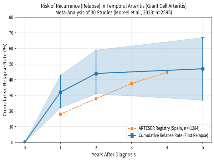
Cumulative relapse risk in temporal arteritis (giant cell arteritis, GCA) rises steeply in the first 1–2 years after diagnosis and then largely plateaus, reaching roughly 45–50% by 5 years in meta-analyses of glucocorticoid-treated patients.
A 2023 systematic review and meta-analysis (Moreel et al.) of 30 studies (2,595 patients) reported these cumulative rates for a first relapse after treatment initiation:
1 year: 32% (95% CI 22–43%)
2 years: 44% (95% CI 31–59%)
5 years: 47% (95% CI 27–67%)
Most relapses occur early (mainly while tapering glucocorticoids, often at doses <10 mg/day prednisone equivalent).
Relapses after year 5 are uncommon.
About 30% of patients have a second relapse and ~17% a third.
Overall, roughly half of patients experience at least one relapse, and about one in three have multiple relapses.
The Spanish ARTESER registry (large real-world cohort) showed somewhat lower early rates that continue rising more gradually (≈18% at 1 year, 28% at 2 years, 38% at 3 years, 45% at 4 years), with most events still concentrated in the first 2 years.
Other cohorts and reviews consistently describe the same pattern: the bulk of risk is front-loaded in the first 2 years, with low incremental risk thereafter under standard care.
Key modifiers
Longer planned glucocorticoid courses are associated with
lower 1-year relapse rates
Female sex and large-vessel involvement increase risk. Tocilizumab (and to a
lesser extent other steroid-sparing agents) substantially reduces relapse rates
during active treatment, though risk often rises again after discontinuation.
The graph below plots the meta-analysis cumulative first-relapse rates (with confidence intervals) and overlays the ARTESER registry points for comparison:
These figures reflect typical glucocorticoid-based
regimens in the literature (often tapered over 12–24+ months).
Individual risk varies with phenotype, treatment
intensity, and monitoring.
Relapse is usually manageable with temporary dose
increases ± steroid-sparing therapy.
Vision-threatening events are uncommon once treatment has begun.
Historical perspective.
  |
Evidence of temporal arteritis: painting by Jan Van Eyck (c. 1385–1440),
In the 15th century, in 1436, it was documented that the Canon Van der Paele of Bruges,a wealthy donor to the church of Saint Donatian in Bruges, was unable to do his daily prayer and comissioned this painting by Van Eyck in response to illness. Clearly visible in the painting are a pair of spectacles, but prominent temporal arteries are clearly visible in the work. Painting and art was the photography of the time. The painting is now in the Groeninge museum Bruges |
|